You are using an out of date browser. It may not display this or other websites correctly.
You should upgrade or use an alternative browser.
You should upgrade or use an alternative browser.
Spot the difference!
- Thread starter Jo90
- Start date
Dr M
Verified Dentist
- Joined
- May 31, 2019
- Messages
- 1,643
- Solutions
- 126
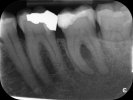
It looks like your second molar on the left has a peri-apical infection. Can't see clearly if the distal root has a fracture as well? More advanced imaging is required, but at the very least a root canal treatment is indicated on this tooth.
Thank you for your reply. Are you referring to the tooth next to the wisdom tooth?
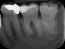
Is that something that only shows on today's X-ray, or did it show on last year's?
I have never had any problems with my teeth apart from once in May 2022 when I suddenly experienced excruciating pain all over the left side of my face. It lasted for about ten days, gradually subsiding. At that time, my dentist couldn't find any cause for it and suggested it could be a result of clenching my teeth in my sleep, although she noted a 4mm pocket in the gum of the lower left tooth next to the wisdom tooth. Since then, I have had no symptoms and no pain, visit my dentist and hygienist regularly, have good oral hygiene and avoid sugary foods. Hence, it was a shock, when she also told me today that I needed root canal work. She said the gum had improved and the tooth was good but there was bacteria in the bone under the tooth that might not even be cured by root canal work. How could this have happened so suddenly; could it be connected to the issue I experienced in May 2022? Is there any alternative treatment?
Dr M
Verified Dentist
- Joined
- May 31, 2019
- Messages
- 1,643
- Solutions
- 126
It sounds like you might have cracked your tooth. I am referring to the tooth next to the wisdom tooth yes. It does not show on the first x-ray, but only on the second x-ray. It might just be that in the first x-ray it was too soon for any changes to appear. A deep pocket might also be an indication of a root fracture. I would suggest maybe asking for a CBCT, just to rule out any fractures. A CBCT provides a 3D image of the tooth. If there is a root fracture,a root canal will be of no use and then the only option would be to extract the tooth and consider replacement options.
The grinding should also be addressed as soon as possible.
The grinding should also be addressed as soon as possible.
- Joined
- Dec 26, 2023
- Messages
- 109
I can report a similar case.
I had cracked the crown of the right tooth (first molar #46) when biting on an almond in early summer of 2023. After filling, it cracked again...then I got 24/7 toothache.
First X-ray taken in early September 2023 (and a CBCT scan, which showed a periapical infection, like yours). Interesting that the X-ray did not show the infection.
The CBCT scan did not show a root fracture, but the endodontist suspected one (root fractures have to have a certain width and the CBCT a certain resolution/field of view to image fractures: this is not always possible).
An oral surgeon in my family confirmed what I read in the literature: once one has a periapical infection, a root canal is required. If there is a fracture, the tooth is lost.
Root canal treatment was initiated in early October, however not completed, as pain did not subside within 11 weeks. The tooth was extracted just before Christmas.
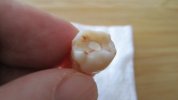
The second X-ray was taken just before the extraction. The oral surgeon in my family interpreted a root fracture (though it may be ambiguous). The root clearly shows the infection (black arrow).
The oral surgeon who extracted the tooth reported a root fracture.
I never had any issues with my teeth before and also have my teeth cleaned every 6 months. The root fracture was assigned to bruxism. I recently received a custom-made nightguard to avoid such fractures in the future.
What I learnt...I can quote from a peer reviewed article: "...They [vertical root fractures] are also difficult to diagnose, as they mimic other conditions. Hence, the diagnosis of vertical root fractures requires more of a predictive rather than a definitive identification. A cumulative assessment of the clinical signs and symptoms and the radiographic features may help us reach a definitive diagnosis..."
Good luck!
P.S. According to the literature I read, a periapical abscess/infection takes 2-10 months to appear radiographically following the pulp infection (inside the root). My dentist spoke of 2-3 months.


I had cracked the crown of the right tooth (first molar #46) when biting on an almond in early summer of 2023. After filling, it cracked again...then I got 24/7 toothache.
First X-ray taken in early September 2023 (and a CBCT scan, which showed a periapical infection, like yours). Interesting that the X-ray did not show the infection.
The CBCT scan did not show a root fracture, but the endodontist suspected one (root fractures have to have a certain width and the CBCT a certain resolution/field of view to image fractures: this is not always possible).
An oral surgeon in my family confirmed what I read in the literature: once one has a periapical infection, a root canal is required. If there is a fracture, the tooth is lost.
Root canal treatment was initiated in early October, however not completed, as pain did not subside within 11 weeks. The tooth was extracted just before Christmas.
The second X-ray was taken just before the extraction. The oral surgeon in my family interpreted a root fracture (though it may be ambiguous). The root clearly shows the infection (black arrow).
The oral surgeon who extracted the tooth reported a root fracture.
I never had any issues with my teeth before and also have my teeth cleaned every 6 months. The root fracture was assigned to bruxism. I recently received a custom-made nightguard to avoid such fractures in the future.
What I learnt...I can quote from a peer reviewed article: "...They [vertical root fractures] are also difficult to diagnose, as they mimic other conditions. Hence, the diagnosis of vertical root fractures requires more of a predictive rather than a definitive identification. A cumulative assessment of the clinical signs and symptoms and the radiographic features may help us reach a definitive diagnosis..."
Good luck!
P.S. According to the literature I read, a periapical abscess/infection takes 2-10 months to appear radiographically following the pulp infection (inside the root). My dentist spoke of 2-3 months.
Last edited:
- Joined
- Dec 26, 2023
- Messages
- 109
Extracted!Dear Brause,
as per the x-ray, visible changes are not there. however if your tooth has become more sensitive or if there is any pain, do consult and Endodontist. the filling is close to the nerve though. For more info you can visit https://urbansmiles.in/
Attachments
Ask a Question
Want to reply to this thread or ask your own question?
You'll need to choose a username for the site, which only take a couple of moments. After that, you can post your question and our members will help you out.
Similar Threads
Forum statistics
Latest Threads
-
Painful Wisdom Tooth Cavity?
- Started by ArcaneWisdom
-
I want to be a omfs
- Started by teba
-
Abscess and amoxicillin
- Started by selenamonet87
-
Could Crowns fix my tooth-surface loss problem?
- Started by william_
-
Should I have a crown?
- Started by Muzza
-
Is this normal appearance for tongue?
- Started by wwwccceee2024
-
Is this normal appearance for tongue? I try to scrape my tongue but it still looks like this
- Started by wwwccceee2024
-
About a cleaning
- Started by iamafraid
-
Fractured crown: tooth 36 to be extracted
- Started by Brause
-
Possible Cut on Gum?
- Started by Chloe_06